 What is oral diagnosis?
Oral diagnosis is operation of establishing diagnosis following the examination of the roentgens belonging to the patient’s oral region and realization of detailed intra-oral examination.
Radiology
The biggest contributor in the diagnosis of the intra-oral diseases – especially the ones related to the hard tissue such as tooth and jawbone – is dental radiographies, that is to say tooth films. Two types of tooth films which are used at most are periapical (showing 2-3 teeth, small) and panoramic (showing all lower and upper teeth and all of jawbones, big) films.
Tooth films (Dental Radiography)
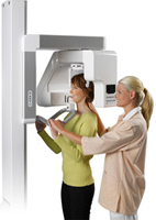
- Digital Panoramic Radiography:
The panoramic radiographies are the technique of imaging of all of the current teeth in the mouth, impacted teeth, bone tissue surrounding the teeth, whole jawbone. While the images belonging to the roentgen drawn can be seen on the computer display immediately, the enlargement-downsizing operations and measurements can be made on these images. In this technique, the rate of radiation to which the patient is exposed has significantly decreased. Also, it is an important advantage that the images can be kept in the patient dossier in the computer environment or can be shared on the internet environment when required. - Periapical radiographies:
It is intra-oral imaging technique which is used with the aim of getting more detailed images regarding the suspicious situations determined in the panoramic radiographies and in which only several teeth which are neighbors to each other and bone tissue surrounding these teeth can be displayed. - Digital radiographies (RVG):
While the images belonging to the roentgen drawn can be seen on the computer display immediately, the color adjustments, enlargement-downsizing operations and measurements can be made on these images. In this technique, the rate of radiation to which the patient is exposed has significantly decreased. Also, it is an important advantage that the images can be kept in the patient dossier in the computer environment or can be shared on the internet environment when required. - Computerized tomography (BT, CT):
It is a three dimensional tomographical imaging technique used in the stations where the traditional intra-oral and extra-oral imaging techniques are insufficient such as big cysts, tumors and cases on which a great number of implant applications are planned. On the contrary to the other two dimensional techniques, latitudinal sections can be taken from the oral region in this method and by this way, the neighborhood of the teeth and pathological formations with the surrounding tissues can be analyzed in 3 dimensions. Especially in the cases where a great number of implants will be applied, the bone thickness in the horizontal direction, locations of the sinus cavities and way that the vein and nerves follow can be determined definitely. One other advantage of this method is that the study models or guide plaques belonging to the requested regions can be prepared before the surgical operation by using as combined with the fast prototyping method. By this way, the risk of the surgical operations has been minimized.